Giới thiệu về kính hiển vi nha khoa trung quốc với máy ảnh
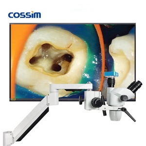
Alibaba.com cung cấp các sản phẩm 967 kính hiển vi nha khoa trung quốc với máy ảnh. Có rất nhiều kính hiển vi nha khoa trung quốc với máy ảnh lựa chọn dành cho bạn, chẳng hạn như oem, odm, và obm. Bạn cũng có thể chọn từ led, led ánh sáng, và halogen kính hiển vi nha khoa trung quốc với máy ảnh. Cũng như từ bằng một mắt, trinocular kính hiển vi nha khoa trung quốc với máy ảnh.Và bất kể kính hiển vi nha khoa trung quốc với máy ảnh là quang học, phòng thí nghiệm nghiên cứu, hay đồ trang sức xác định.